Medical translation requested
Posted: Mon Mar 30, 2015 10:54 am
I've been having numbness in my hands, starting to creep up my forearms, for about a month and a half. My doctor, suspecting nerve involvement, ordered an MRI. The report is in the spoiler below:
My doctor (actually, the doctor covering for him) was sufficiently concerned when she saw this report Thursday night to put in what she called an "urgent referral" to a surgeon, which she said I can expect to occur this week.
I'm pretty sure I get the gist of what's going on, but if one of the medical professionals would be kind enough to translate for me, I'd appreciate it. If someone can give me an idea of what to expect when I get my referral, that would be doubly appreciated.
Thanks in advance. --Bob
Long technical document
March 22, 2015
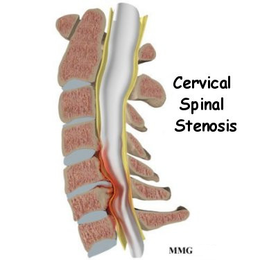
MRI OF THE CERVICAL SPINE:
HISTORY: neck pain with bilateral arm numbness/burning x 3 weeks
COMPARISON: None.
TECHNIQUE: On a 3 Tesla scanner, sagittal T1-weighted, T2-weighted and STIR images and axial T1-weighted and T2-weighted images were obtained.
FINDINGS:
There is reversal of the normal cervical lordosis. Vertebral body heights are normal except for mild degenerative height loss in the mid cervical spine. There is a grade 1 anterolisthesis at C3-C4 due to facet degeneration and mild degenerative retrolisthesis at C4-C5 and C5-C6 due to disc degeneration. Discogenic degenerative endplate changes are noted adjacent to multiple intervertebral discs.
There is moderate degenerative disc narrowing from C4-C5 through C6-C7 and mild degenerative disc narrowing in the upper cervical spine.
At C2-C3, there is mild disc bulging without significant spinal canal stenosis. There is moderate left foraminal narrowing due to left facet osteophytes. There is no significant right foraminal stenosis.
At C3-C4, there is circumferential disc bulging and ligamentum flavum/facet degeneration, causing moderate spinal canal stenosis and flattening of the dorsal and ventral surfaces of the spinal cord. There is moderate bilateral facet degeneration with multiple small subchondral cysts on the left. There are also edematous changes in the marrow of the articular processes. There is severe bilateral neural foraminal stenosis related to the anterolisthesis and disc/osteophyte complex.
At C4-C5, there is a circumferential disc bulge/osteophyte complex, mildly narrowing the spinal canal and flattening the ventral spinal cord. There is moderate bilateral foraminal narrowing due to the disc/osteophyte complex.
At C5-C6, there is a circumferential disc bulge/osteophyte complex, mildly narrowing the spinal canal. There is mild to moderate bilateral foraminal narrowing due to the disc/osteophyte complex.
At C6-C7, there is a small circumferential disc/osteophyte complex without significant spinal canal stenosis. There is mild bilateral foraminal narrowing due to the disc/osteophyte complex.
There is no mass or hematoma in the spinal canal. There is abnormal T2-weighted hyperintensity and likely volume loss in the spinal cord at the C3-C4 level.
The paraspinal soft tissues are within normal limits.
IMPRESSION:
Moderate multilevel disc and facet degeneration, as described above.
Moderate spinal canal stenosis at the C3-C4 level related to disc bulging, facet degeneration, and the resulting anterolisthesis. There is also abnormal T2-weighted hyperintensity in the spinal cord at this level that likely represents spinal cord myelomalacia.
Severe bilateral neural foraminal narrowing at C3-C4 and milder degrees of foraminal narrowing at multiple additional levels, as described above.
I'm pretty sure I get the gist of what's going on, but if one of the medical professionals would be kind enough to translate for me, I'd appreciate it. If someone can give me an idea of what to expect when I get my referral, that would be doubly appreciated.
Thanks in advance. --Bob